Saturday 8 September 2018

The Future of Neuroscience


In the current animal model of depression, a mouse is placed in a jar of water and struggles to swim to avoid drowning, in the aptly named “forced-swim” test(1). After a few minutes, the mouse stops trying to escape, instead choosing to float immobile in the water. At this point, the mouse is said to experience “behavioural despair” (the mouse loses hope to escape the stressful environment) and the mouse is then classified as suffering from “depression”. This is the standard model used to test antidepressant drugs – the time spent immobile versus swimming in mice given the drug is compared to that of controls. Clearly, this is a simplistic model with very little resemblance to the highly complex, multi-faceted disorder of clinical depression in humans. Yet, although its efficacy has long been contested(2), this is still the most common mouse model used in the majority of research into “depression”. 

This is not just the case for depression. Many psychiatric disorders are still studied using simplistic animal models which – though important – essentially bear little resemblance to the experiences of those suffering from the disease on a daily basis. 

On top of this there, is a pervasive disconnect between psychology and neuroscience. It is fundamentally impossible to study affective or cognitive phenomena such as emotion or foresight using animal models, since mice, rats and monkeys are all unable to communicate what they are feeling with humans. Instead, researchers study behaviours as a proxy – for instance, what a mouse does before it gets a reward, which is largely a matter of interpretation. But we lack a way to study actual emotions in mice – which, besides, are likely vastly different subjective experiences to those of humans. While mouse models have their uses, they are generally an insufficient representation of brain disease or even normal brain function in humans. 

It is without surprise then, that over the past 40 years there has been little improvements in the outcomes of patients with the most common brain diseases. Some pharmaceutical companies are abandoning research into drugs for psychiatric diseases altogether due to the high cost and low success rate. For example for Alzheimer’s, every time we think we have a promising new drug in development to break down the toxic amyloid plaques, we find that it fails in clinical trials, and moreover, we find that we were coming at the problem from the wrong angle altogether(3). We now know that we need to intervene long before amyloid deposits are prevalent, and long before symptoms are seen. Some research shows a portion of patients diagnosed with Alzheimer’s do not even have significantly more amyloid-β plaques in their brains than healthy controls, and amyloid pathology has been observed in cognitively healthy elderly individuals, suggesting that amyloid-driven tauopathy may at best only be part of the problem. Thus, we currently lack even an effective diagnostic criteria for neurodegenerative disorders. Treatment for Alzheimer’s is largely symptomatic; we are a long way off from understanding the root causes of the disease. Progress is slow, but we are learning. 

There is also the problem of brain scanning. The most prevalent form of brain imaging in neuroscience and psychology is undoubtedly functional magnetic resonance imaging (fMRI). However, again, this is a proxy – fMRI measures blood flow across the brain while the subject is engaged in a particular task or activity; it does not directly measure neuronal activity(4). One unpublished study from 2009 found apparent cognitive activity in the brain of a dead salmon(5), highlighting the risk of false positives in fMRI studies. Similarly, electroencephalography (EEG) measures electrical activity at the brain surface – however it lacks specificity in that it does not measure the activity of specific neurons or sets of neurons, but rather of a crude combination of electrical currents across a particular brain area. Unfortunately, it is not yet possible to measure the activity of a specific set of neurons in living, human brain tissue. 

However, a small minority of forward-thinking neuroengineers are currently working on measuring real-time electrical brain activity in vivo, in humans. This is already possible in the brains of mice and in monkeys, but not yet in humans. Thus, hopefully in the not-too-distant future, we will be able to record activity from specific sub-sets of neurons and correlate this with not only behaviour, but with thought, emotion and, of course, depression. 

With a little imagination, let’s fast forward 100, or perhaps only 50 years. We now understand the root causes of Alzheimer’s, Parkinson’s, depression, schizophrenia etc., and are able to deliver targeted genetic or drug therapies, custom-made for each patient, to treat brain disease both symptomatically, and more importantly, prophylactically. Furthermore, we now understand that these disorders which we considered one disease, were in fact different diseases with similar symptoms but vastly different biological causes, each requiring a different treatment. We will look back to the primitive days of neuroscience – the early 21st century – and be amazed that the majority of brain diseases were being treated with the wrong drugs, which were more often than not completely ineffective, or even counter-productive(6,7).

In order to achieve this, we first had to figure out how to get electrodes through the skull and into the brains of healthy, living humans, without causing any risk to the subject. Rather than drilling holes through the skull, we use microelectrodes so small that they can be inserted without rupturing any blood vessels, thus avoiding the risk of stroke. We might even use lasers. As technology progresses, we will be able to record from thousands of electrodes at once using smart, robotic, microscopic implantations which work their way around blood vessels and through the brain tissue. Eventually, this will be possible using wearable devices which the subject can implant into their brain and go about their day, while the device is constantly collecting and uploading high-resolution neuronal activity directly from their brain to the computer of a researcher, or their doctor, for analysis. Combined with powerful yet harmless lasers able to pass through the skull and produce images of neurons and synapses with sub-cellular precision, we are able to decipher not only the connectome, but the precise patterns of activity between specific neurons during a particular function, on an individual level, for each patient. By collecting masses of data from millions of patients, we can mathematically calculate – with the aid of superfast computers – what exactly is going wrong in neurological diseases such as Alzheimer’s. Furthermore, we have finally managed to bridge the gap between psychology and neuroscience, by being able to ask the patients about their emotional, subjective experiences, and correlate this with their neuronal activity at that exact point in time. 

At some point, these wearable devices will become commercialised by the likes of Google, Amazon and Apple, offering free services to customers in exchange for their private data – their thoughts. Having learned from our mistakes in the early 21st century regarding privacy and data harvesting, customers will demand rights and legislation to decide how their personal brain activity is used by multinational corporations. However, this will prove ineffective, and customers will willingly sacrifice their privacy anyway, by updating to the latest version of Apple iBrain® without reading the terms and conditions. This will open up a whole Pandora’s box of neuro-hacking and neuro-spyware, as well as further driving inequality and elitism – since only those in first-world countries can access the devices, and only the wealthiest of those can afford the latest and greatest bio-upgrades. The societal, political and economic ramifications of this could fill an entire book in and of itself. But from a neuroscientific perspective, this will be a turning-point; a revolution in neuroscience research, allowing for not only the enhancement of normal brain function – or biohacking – but also significant advances in the treatment of brain diseases. Alzheimer’s, schizophrenia, autism, ADHD, addiction, depression and anxiety disorders will all be things of the past. 

So too will smartphones. Generation Y will tell their kids, “I remember when we had to type our text messages with our thumbs, or ask Alexa to add vegan meat to the shopping list. We never had Google Think® in my day”, or “I remember when we had to go to college to study for years, we had to sit down and read books to learn things. We never had Amazon HiveMind® in my day”. Meanwhile their kids seamlessly communicate via Apple iThought®, video chat via Skype Hologram®, and instantaneously download entire textbooks and literature via an ultra-fast 100Gb/s subscription to Amazon’s entire library for only $19.99 per month. Fake news will become a thing of the past, as every news article you download is instantly verified against thousands of peer-reviewed sources – reviewed both by humans and by sophisticated AI technology. You will never forget anything ever again, as any memory you choose to remember will be uploaded to the cloud, ready to be accessed and relived at will. Alternatively, should you choose, you can delete a traumatic or stressful memory, like it never happened. Without delving too far into the realm of science fiction, the possibilities are Limitless®. Anything is possible, so long as we can dream it – or Google DeepDream® it. 

Our knowledge of neuroscience is only in its infancy. Our understanding the human brain in all its complexity is only a mere few steps away from exponential growth. As technology combines with neuroscience, we become ever closer to understanding ourselves, and to an entirely interconnected consciousness. Societies working together as a collective intelligence are capable of amazing things – just look at bees and ants. Times are changing, for better or for worse.

Now, back to those mice...


References: 

1. Can, Adem, Dao, David T., Arad, Michal, Terrillion, Chantelle E., et al. (2012) ‘The Mouse Forced Swim Test’. Journal of Visualized Experiments : JoVE, (59). [online] Available from: https://www.ncbi.nlm.nih.gov/pmc/articles/PMC3353513/
2. Borsini, Franco, Volterra, Giovanna and Meli, Alberto (1986) ‘Does the behavioral “despair” test measure “despair”?’ Physiology & Behavior, 38(3), pp. 385–386.
3. Castello, Michael A., Jeppson, John David and Soriano, Salvador (2014) ‘Moving beyond anti-amyloid therapy for the prevention and treatment of Alzheimer’s disease’. BMC Neurology, 14, p. 169.
4. Ekstrom, Arne (2010) ‘How and when the fMRI BOLD signal relates to underlying neural activity: The danger in dissociation’. Brain Research Reviews, 62(2), pp. 233–244.
5. Scicurious (2012) ‘IgNobel Prize in Neuroscience: The dead salmon study’. Scientific American Blog Network. [online] Available from: http://blogs.scientificamerican.com/scicurious-brain/ignobel-prize-in-neuroscience-the-dead-salmon-study/ (Accessed 27 April 2016)
6. Anon (2016) ‘Most antidepressant drugs ineffective for children and teens, study finds’. University of Oxford. [online] Available from: http://www.ox.ac.uk/news/2016-06-08-most-antidepressant-drugs-ineffective-children-and-teens-study-finds (Accessed 6 July 2018)
7. Cipriani, Andrea, Zhou, Xinyu, Giovane, Cinzia Del, Hetrick, Sarah E., et al. (2016) ‘Comparative efficacy and tolerability of antidepressants for major depressive disorder in children and adolescents: a network meta-analysis’. The Lancet, 388(10047), pp. 881–890. 

Friday 6 July 2018

Forgotten Memories Brought Back in Mice? Hold Up...

A study published yesterday in Cell[1] has purportedly found that memories formed as infants may be able to be retrieved as adults using optogenetic techniques, and various media outlets have enthusiastically inferred that this suggests we may be able to remember the events from our infancy, some even suggesting we may be able to remember our own birth. 

The study, led by psychologist Paul Frankland, was based on previous research by the group which found that the “forgetting” of memories during infancy may be a result of high levels of hippocampal neurogenesis at this age – i.e. the neurons representing the memories are replaced by new neurons, thus erasing the memories. 

The aim of the current study was to determine if the memories formed during infancy are permanently lost due to a failure in encoding during infancy, or become progressively inaccessible over time due to a progressive loss in the ability to retrieve them as the mice age. 

The researchers placed young mice into a box and gave them a foot-shock, so that when they are placed back into the box they freeze in anticipation of the shock - a classic fear-based training paradigm. The mice had been engineered to contain a specific set of light-sensitive neurons in a particular region of the hippocampus involved in the formation of memories - the dentate gyrus - which allowed the researchers to activate these neurons by firing a laser at them. They then placed the same mice back into the box as adults and activated this set of neurons, thus reinstating the memory and causing the mice to freeze in anticipation of the shock. 

The authors seem to suggest that this means that some hidden traces of the memories created during infancy were retained and were able to be recalled by the researchers by optogenetically activating the specific set of hippocampal neurons which were observed to be activated during the contextual fear encoding (the “dentate gyrus encoding ensemble”), positing that the reactivation of these ensembles was sufficient for “memory recovery” in adulthood. 

They then quantified the activity of an activity-regulated gene, c-Fos, in cortical and subcortical brain regions following the fear learning event. The purpose of this was to determine whether fluorescently-tagged neurons activated during the memory encoding event were preferentially reactivated during the “memory recall” in adulthood – which of course they were, implying successful recall of the memory. 

However, while they may have been able to reactivate a neural pathway they have essentially programmed into the mice’s brains during infancy, I’m not so sure it was the actual memories themselves which were “recovered”. 

It’s worth nothing that these are memories which the researchers have created in the mice by giving them foot-shocks within a particular environmental context; they are not naturally formed memories. 

“When the infant mice were placed in the box and the laser was turned on, the animals’ memories of the electric shock returned and they froze in place.” 

The fact that the mice freeze when they are placed into the same environment in which they were given a foot-shock during infancy does not necessarily mean that they remember receiving the foot-shock. It simply means that a particular behaviour (freezing) has been programmed into their brains and artificially re-instated by firing lasers at the neurons underlying this behaviour. It means that the same neural pathways resulting in a fear-based freezing response were activated – but these may be totally separate from the pathways containing the actual memory of the event (if they even exist). Besides, we are talking about a simple, conditioned response here; an instinctual behaviour in response to pain – much like you learn to quickly move your hand away from a hot plate – not an actual subjective, detailed memory of an event. It’s possible that the mice would freeze if placed in an entirely different context and the same light-sensitive neurons artificially reactivated.

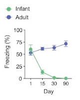
“To first induce memory formation in the animals, the scientists placed the mice in a box and gave them a mild foot shock. While young adult mice retained this memory and froze when put in the box a second time, infant mice forgot this fear-related memory after a day and behaved normally when they encountered the box again” 
Percent freezing levels declined with retention delay in P17, but
not P60, mice. From Guskjolen et al., 2018, Figure 1(B).

So the infant mice were not forming the memories? This appears to contradict the conclusions of the study – i.e. that those memories are simply hard-to-retrieve; hidden deep within the brain, unable to be recovered by natural cues, and that direct stimulation of the engram (in combination with re-exposure to the training context) may reinstate the connections, leading to memory recovery. How can we say that the memories are simply difficult to retrieve when we are unsure if they were ever formed in the first place? 

“However, we found that opto-stimulation of neural ensembles that were engaged during training was sufficient to induce conditioned freezing at the same retention delays. These results suggest that the underlying engram corresponding to the fear conditioning event is not completely overwritten. Rather, this engram presumably exists in an otherwise inaccessible, dormant state, in which “natural” reminders (such as exposure to the training context) most often do not induce successful reactivation(...) This pattern of results is reminiscent of other amnestic states, including mouse models of retrograde amnesia and Alzheimer’s disease, in which opto-stimulation of tagged encoding ensembles (but not presentation of natural cues alone) permits memory recovery." 

Again, the conclusions drawn assume that the artificially activated ensembles encode the actual memory of the event itself – which is not only not confirmed, but hard to believe considering when the mice were put back in the box the second-time as infants, they had not remembered the fear-related memory supposedly created the day before. How, then, do we know that the memory was encoded at all? How do we know it is the memory that is recalled, and not simply a programmed, artificially instated fear-response? This is too simplistic of a model to draw such far-fetched conclusions, and we certainly can’t say that this type of “forgetting” in infancy is akin to other types of amnesia such as that in Alzheimer’s disease. They are completely different processes, at completely different ages. 

Furthermore, less cortical “re-engagement” was observed following optogenetic stimulation of the dentate gyrus engrams in mice trained as infants compared to those trained as adults, further highlighting the possibility that the memories which were purportedly retrieved may not have been formed at all in the infants. 

“Indeed, whereas adult contextual fear memories are successfully consolidated over the course of weeks, equivalent infant memories are being actively forgotten during this period and therefore perhaps not successfully consolidated in the cortex (…) opto-stimulation of tagged dentate gyrus ensembles leads to recovery of an engram that is qualitatively different (and likely impoverished) compared to the equivalent representation in adult animals.” 

The authors even concede that the “memory recovery” did not persist into the light OFF periods – i.e. when the trained mice were placed into the box as adults, they did not freeze unless the hippocampal engrams engineered to be light-sensitive were activated by the researchers – a pattern which has been observed in similar studies involving reactivation of tagged engram cells in the dentate gyrus [2–7]

While the study further adds weight to the idea that infantile forgetting is likely due to a failure of memory encoding in the infant brain (something which we knew anyway), the methods used are simply insufficient to be able to draw some of the conclusions the authors propose, and the study certainly does not suggest that we may be able to recover our infantile memories anytime soon.


 References: 

[1] A. Guskjolen, J.W. Kenney, J. de la Parra, B.A. Yeung, S.A. Josselyn, P.W. Frankland, Recovery of “Lost” Infant Memories in Mice, Current Biology. 0 (2018). doi:10.1016/j.cub.2018.05.059.
[2] X. Liu, S. Ramirez, P.T. Pang, C.B. Puryear, A. Govindarajan, K. Deisseroth, S. Tonegawa, Optogenetic stimulation of a hippocampal engram activates fear memory recall, Nature. 484 (2012) 381–385. doi:10.1038/nature11028.
[3] T. Kitamura, S.K. Ogawa, D.S. Roy, T. Okuyama, M.D. Morrissey, L.M. Smith, R.L. Redondo, S. Tonegawa, Engrams and circuits crucial for systems consolidation of a memory, Science. 356 (2017) 73–78. doi:10.1126/science.aam6808.
[4] D.S. Roy, S. Muralidhar, L.M. Smith, S. Tonegawa, Silent memory engrams as the basis for retrograde amnesia, Proc. Natl. Acad. Sci. U.S.A. 114 (2017) E9972–E9979. doi:10.1073/pnas.1714248114.
[5] T.J. Ryan, D.S. Roy, M. Pignatelli, A. Arons, S. Tonegawa, Memory. Engram cells retain memory under retrograde amnesia, Science. 348 (2015) 1007–1013. doi:10.1126/science.aaa5542.
[6] D.S. Roy, A. Arons, T.I. Mitchell, M. Pignatelli, T.J. Ryan, S. Tonegawa, Memory retrieval by activating engram cells in mouse models of early Alzheimer’s disease, Nature. 531 (2016) 508–512. doi:10.1038/nature17172.
[7] S. Ramirez, X. Liu, P.-A. Lin, J. Suh, M. Pignatelli, R.L. Redondo, T.J. Ryan, S. Tonegawa, Creating a false memory in the hippocampus, Science. 341 (2013) 387–391. doi:10.1126/science.1239073.

Wednesday 3 January 2018

The role of epigenetics in the neurobiology of stress and psychiatric diseases


Epigenetics refers to potentially heritable, experience-dependent molecular alterations to regulation of genetic function, or to numerous proteins or transcriptional regulators that alter the expression of genes, without altering the underlying DNA code itself – in other words, “nuclear inheritance which is not based on differences in DNA sequence"(1).

Figure 1: (a) Structure of eukaryotic chromatin.
(b) Known post-translational modifications. From Sun (2013).
Since the genome contains a massive amount of genetic information which must be condensed into a small space, nuclear DNA is found bound to small, basic, lysine-rich proteins called histones which serve to package the DNA into tightly compact units called nucleosomes. The histone octamer consists of two of each of the four core histone proteins H2A, H2B, H3 and H4, as well as H1 linker histones. Each of these have protruding N-termini tails which are subject to various post-translational modifications such as acetylation, methylation and phosphorylation. These modifications recruit various proteins, including histone modifiers, transcription complexes etc., which alter the chromatin, thereby dynamically altering its function(2). 

There are hundreds of such modifications which act together in complex combinations. Some of the most common are acetylation, methylation and phosphorylation. Acetylation is thought to activate gene transcription by neutralising the histone molecule’s positive charge, thereby disrupting its interaction with the negatively charged DNA, resulting in a remodelling of the chromatin into a more “relaxed” structure which is more accessible to transcription factors(3). Acetyl groups are added to lysine by a group of enzymes called histone acetyltransferases (HATs), and removed by histone deacetylases (HDACs)4. Similarly, methylation involves the addition of methyl groups to lysine residues. Addition of a methyl group by a histone methyltransferase (HMT) generates a docking site which recruits transcriptional regulators to specific gene loci4. This can either activate or repress transcription depending on the specific residue involved. DNA itself can also be methylated, repressing expression by interfering with the binding of transcription factors(4). Conversely, histone phosphorylation is generally associated with transcriptional activation.

These epigenetic modifications, along with plasticity, play a crucial role in many aspects of stress pathology including hypothalamo–pituitary–adrenocortical (HPA) axis function, reactivity to glucocorticoids in psychiatric diseases such as PTSD and anxiety, and the disease pathology of depression(5). Thus, epigenetics is a mechanism by which life experiences transduce genetic changes which, combined with existing genetic predispositions to psychiatric diseases such as depression, addiction and schizophrenia, can alter an individuals vulnerability to expressing the disease phenotype.

There are two main categories of animal models used to study depression. The first involves placing the animal under acute stress, such as the forced swim and tail suspension tests; the second involves exposing the animal to chronic stressful stimuli – such as in the chronic mild stress (CMS), chronic unpredictable stress (CUS) or chronic social defeat models.

The CMS model involves continuously exposing mice to a range of mild stressors, such as food/water deprivation, temperature reductions, restraint stress etc., which over a period of weeks leads to gradual reduction of preference for sucrose – an indication of anhedonia, a hallmark symptom of depression – which is reversed by chronic treatment with antidepressant drugs(6). The CUS model is similar except that the stressors are delivered randomly, at unpredictable times.

In the social defeat model, mice are repeatedly confronted with an aggressive male mouse for a period of 30 days, placing them under psycho-emotional stress and leading to the development of anxious-depressive symptoms as determined by behavioural tests(7). Defeated mice display social avoidance when subsequently confronted with an unfamiliar mouse – a characteristic symptom of depression – as compared to controls(8). Mice placed under chronic stress also show anhedonic symptoms such as a decrease in reward behaviours such as preference for sucrose(8). These depressive-like symptoms are also seen in the offspring of defeated mice(9), and neurodevelopmental deficits in mice bred from stressed mothers have even been characterised in up to third generation mice(10). Male mice exposed to social defeat or maternal separation – separating pups from their mother, another early life stress model which leads to depression-like behaviours – produce offspring which are also more vulnerable to stress(11). This stress-vulnerability in turn may be epigenetically transmitted to their offspring by changes in DNA methylations states, and thus changes in expression of, certain genes in their germ cells(12).

Furthermore, the offspring of rats which exhibit high levels of licking/grooming (LG) show significantly less anxiety/depression-like symptoms than those from mothers which exhibit low levels of LG behaviour(13), and are less reactive to stress in later life(14). Analysis of neuroendocrine levels of the offspring of low LG rats vs. high LG rats reveals distinct differences in HPA axis function and glucocorticoid mRNA expression(15). Additionally, persistent differences in DNA methylation at certain regions of the genome are observed between low LG and high LG rats throughout later life(14), and CUS mice have been shown to have increased activity of the histone deacetylase HDAC2(16). Moreover, female rodents raised by less nurturing mothers in turn show less nurturing behaviour towards their own offspring, as well as higher anxiety levels(17) – a phenomenon also seen in humans.

However, not all mice exhibit the same vulnerability to such stress-induced epigenetic changes. About a third of mice display a resilience to developing depression-like behaviours after exposure to the same stressors, and these mice show differences in levels of specific epigenetic changes – including DNA methylation and histone acetylation/methylation – in specific brain regions, particularly those involved in reward pathways(11). By blocking specific epigenetic modifications, these mouse models can be used to effectively study the importance of these modifications in the development of stress-induced pathologies, with the aim of developing more effective treatments for psychiatric diseases such as depression.

Depression is the result of a disorder of many different circuits within the brain, and as such shows a high level of heterogeneity. Many epigenetic changes have been identified in animal models which lead to the expression of depression-like behaviours in rodents, or resilience to the development of such behaviours. There is increasing evidence that epigenetic changes are also important in the pathogenesis of depression in humans, with similar mechanisms being identified in post-mortem brain samples of depressed humans(2). Depression is known to be heritable – genetics has been estimated to account for ~40% of the risk(18). This is, however, considerably lower than the heritability of other psychiatric diseases such as schizophrenia(2), which combined with the high discordance rate between monozygotic twins (50%)(19), suggests that other factors – such as epigenetics – are involved. In fact, monozygotic twins show significantly increased differences in levels of DNA methylation and histone acetylation as they grow older(19). Moreover, it has been suggested that epigenetic changes may be passed down to the offspring of affected individuals, causing changes in HPA axis function, vasopressin and serotonergic systems and increasing their vulnerability to depression(9,20). Mice subjected to unpredictable maternal separation (MS) who showed increased depression-like symptoms indicated by increased time spent floating in the forced swim test (a measure of helplessness) showed significant improvement when administered antidepressant desipramine vs. saline, as expected. However, interestingly, the depression-like symptoms were also observed in the offspring of MS mice who were reared normally, as well as third-generation mice(12) – suggesting epigenetic changes, to some extent, may be heritable.


Epigenetic changes (methylation) have been identified in glucocorticoid genes important for HPA axis function in the hippocampi of human victims of childhood trauma compared to those with normal childhoods(21,22), and decreased expression of brain-derived neurotrophic factor (BDNF) has been observed in post-mortem brain samples of depressed patients(23) compared to healthy controls – significant since changes in BDNF expression are required for the development of social avoidance induced by social defeat, and knockdown of BDNF expression in the nucleus accumbens (NAc) blocks this by the same mechanism as chronic treatment with antidepressants(8). Furthermore, adaptations within mesolimbic dopamine pathways, of which the NAc is a key component, have been identified as underlying the susceptibility or resilience of mice to develop depressive symptoms as a result of social defeat, specifically anhedonia(24). Susceptibility to depression induced by chronic social defeat has been associated with decreased levels of several histone methyltransferases (HMTs) – including G9a – in the NAc of susceptible mice, while these HMTs are upregulated in resilient mice(25). Additionally, artificial knockdown of G9a was associated with increased susceptibility to depression-like behaviours. Global increases in methylation of H3K4 residues has also been identified as a mechanism of non-selective antidepressant drugs (monoamine oxidase inhibitors), as a result of inhibition of H3K4 demethylation in the nucleosome, leading to alterations in gene expression(26). More recently, decreased expression of another histone demethylase in the NAc, JMJD2, has also been observed in mice subjected to chronic social defeat, and blocking of histone demethylase using inhibitors resulted in depression-like phenotype even in the absence of stress exposure, suggesting that activation of these enzymes may be an effective target for pharmacological treatment of depression(27).

As well as methylation, histone acetylation in the NAc has also been identified as an important epigenetic factor. Chronic social defeat causes a persistent increase in H3 acetylation in mice, which is associated with reduced levels of histone deacetylase 2 (HDAC2) in the NAc; reduced levels of HDAC2 has also been observed in post-mortem NAc samples from depressed humans(28). Additionally, another histone deacetylase, HDAC5, is associated with resilience, and chronic antidepressant treatment effectively increased expression of HDAC5, resulting in a decreased susceptibility to depression(29). This effect is however specific to the NAc, as HDAC5 administered virally into the hippocampus reversed the drug’s antidepressant effect(30). Thus, specific epigenetic modifications can have varying effects dependent on the brain regions involved.

Changes in expression of transcription factors have also been implicated. ΔFosB, which persists for long periods of times once expressed, can act as both an activator of transcription and a repressor(31). ΔFosB has been shown to be required for the commonly prescribed antidepressant fluoxetine to reverse depression-like behaviours induced by social defeat in mice, similarly to BDNF(32).


As well as being important in the aetiology of depression, the transcription factor ΔFosB has also been shown to act as a “molecular switch” for addiction(33). Following repeated administration of many different drugs of abuse, increased levels of ΔFosB is seen in the NAc, as well as in the dorsal striatum, another region involved in reward pathways. ΔFosB is thought to initiate and maintain changes in gene expression long after drug administration has ceased, perhaps underlying the risk of relapse. Overexpression of the transcription factor causes increased sensitivity to the drug – associated with increased likelihood of addiction – as well as increased drug-seeking behaviour(33). Once initiated, ΔFosB represses the expression of G9a in the NAc, causing increased dendritic spine plasticity of NAc neurons – a hallmark feature of addiction(34,35). Similar to in depression, this epigenetic reduction of G9a induced by cocaine exposure causes a global reduction in H3K9 methylation, which is thought to contribute to the potentiated behavioural responses to cocaine(4), and the subsequent establishment of addiction(36). When G9a is artificially overexpressed in the NAc, cocaine-addicted mice become less susceptible to stress compared to wild-type addicted mice in which G9a is reduced(25). Thus, G9a effectively serves as a molecular marker of resilience. This also suggests overlapping epigenetic mechanisms in the increased susceptibility to stress caused by depression and addiction.

FosB, as well as c-fos genes in the striatum, have been found to show an increased acetylation at histone H4 subunits(37). Activation of these genes by acetylation of the promoter regions, induced by chronic exposure to cocaine, appears to play a long-term role in addiction – indeed, these genes show increased acetylation for days to weeks after the last drug exposure(4). It is thought that chronic cocaine exposure achieves this by increasing the phosphorylation of HDAC5 causing it to be removed from the nucleus, thus allowing histone H4 to be acetylated by HATs at these genes(29). This is supported by evidence that knockdown of the HAT, or overexpression of HDACs, results in a reduced sensitivity to cocaine(29).

Schizophrenia is another psychiatric disease which has been shown to be regulated by epigenetic mechanisms. A common mouse model of schizophrenia is the methylazoxymethanol (MAM) model, in which H3K9 acetylation is decreased due to an increase of histone deacetylase 2 (HDAC2), leading to neurobehavioral deficits characteristic of schizophrenia. Recent experiments using this model have shown that valproic acid (VPA), a short chain fatty acid often prescribed for its mood-stabilising effects, prevents this increase in HDAC2, inducing increased acetylation at specific gene promoters implicated in the pathology of schizophrenia(38,39). Another drug commonly prescribed in the treatment of schizophrenia is the antipsychotic clozapine – a D2 receptor antagonist. Clozapine, however, shows higher therapeutic efficacy compared to other D2 receptor antagonists(40), and it is thought that this may be in part due to its additional ability to upregulate methylation of H3K4 residues at the Gad1 gene promoter, which is crucial in the synthesis of GABA(41). Reduced expression of Gad1 in the prefrontal cortex has been identified in the brains of some schizophrenic patients(42); thus, methylation of H3K4 residues at the promoter region of this gene may explain the enhanced efficacy of clozapine in the treatment of psychosis/schizophrenia.
As well as DNA/histone modifications, RNA can also be subject to epigenetic changes which can play a role in disease pathology. A recent study using RNA sequencing found a significant down-regulation of the long non-coding RNA (lncRNA) Gomafu, which is thought to play a role in anxiety as well as the expression of schizophrenia-related genes(43).

Epigenetic mechanisms are thus increasingly becoming recognised as important factors in the pathology of many psychiatric diseases including depression, addiction and schizophrenia. Drugs which target key enzymes which modify histones/chromatin, such as histone deacetylase inhibitors, may prove effective in the treatment of depression – an area in which novel drug targets are lacking.

However, there are some difficulties when studying epigenetic mechanisms. For example, although depression-like behaviours have been shown to be transmitted across generations, if in vitro fertilisation (IVF) is used in such mice models – i.e. artificially impregnating female mice with sperm from socially defeated mice – susceptibility for depression-like behaviours is not significantly increased in the offspring(44). It would be interesting to investigate why this is – perhaps pre-natal environment plays a key role in the transmission of epigenetic changes induced in the parent mice. More studies using IVF would help to elucidate this.

Additionally, due to the high diversity of the human genome, a great number of subjects will need to be studied in order to obtain accurate data. Genome-wide association studies (GWAS) could be useful. Additionally, epigenetic changes in RNA could be far more important than hitherto recognised. The use of RNA sequencing, similar to Spadaro et al (43), to identify the relevance of long non-coding RNAs (lncRNAs) in the regulation of epigenetic processes relevant in the development of psychiatric disorders could also pave the way for the development of novel drug targets.

Furthermore, researchers often study epigenetic modifications by altering the expression, or levels, of certain enzymes such as HATs, HDACs, or HMTs. However these enzymes are non-specific in that they affect thousands of genes. Thus, it is vital to establish reliable methods to target one specific type of modification at a specific gene promoter, in specific cell types – perhaps combined with GWAS – in order to produce more useful and reliable data.

A final important factor to consider in mouse models is that in some cases, the stress imposed on the adult mice may cause them to interact with their pups differently, which in turn may cause the behavioural changes seen in the pups – rather than epigenetic mechanisms underlying the behavioural differences being transmitted genetically. While these behavioural changes may be epigenetic in nature, it is important to distinguish these from those inherited directly from the parent.

The importance of epigenetic factors in the pathology of psychiatric and neurological diseases is only recently being discovered, and continues to be a key area of study in modern neuroscience research.


References


(1) Wu, C. T., and Morris, J. R. (2001) Genes, Genetics, and Epigenetics: A Correspondence. Science 293, 1103.
(2) Sun, H., Kennedy, P. J., and Nestler, E. J. (2013) Epigenetics of the Depressed Brain: Role of Histone Acetylation and Methylation. Neuropsychopharmacology 38, 124–137.
(3) Kouzarides, T. (2007) Chromatin Modifications and Their Function. Cell 128, 693–705.
(4) Renthal, W., and Nestler, E. J. (2009) Chromatin regulation in drug addiction and depression. Dialogues Clin Neurosci 11, 257–268.
(5) Radley, J. J., Kabbaj, M., Jacobson, L., Heydendael, W., Yehuda, R., and Herman, J. P. (2011) Stress risk factors and stress-related pathology: Neuroplasticity, epigenetics and endophenotypes. Stress 14, 481–497.
(6) Willner, P. (2005) Chronic Mild Stress (CMS) Revisited: Consistency and Behavioural-Neurobiological Concordance in the Effects of CMS. Neuropsychobiology 52, 90–110.
(7) Avgustinovich, D. F., Kovalenko, I. L., and Kudryavtseva, N. N. (2005) A Model of Anxious Depression: Persistence of Behavioral Pathology. Neuroscience and Behavioral Physiology 35, 917–924.
(8) Berton, O., McClung, C. A., DiLeone, R. J., Krishnan, V., Renthal, W., Russo, S. J., Graham, D., Tsankova, N. M., Bolanos, C. A., Rios, M., Monteggia, L. M., Self, D. W., and Nestler, E. J. (2006) Essential Role of BDNF in the Mesolimbic Dopamine Pathway in Social Defeat Stress. Science 311, 864.
(9) Dietz, D. M., LaPlant, Q., Watts, E. L., Hodes, G. E., Russo, S. J., Feng, J., Oosting, R. S., Vialou, V., and Nestler, E. J. (2011) Paternal Transmission of Stress-Induced Pathologies. Biological Psychiatry 70, 408–414.
(10) Dunn, G. A., Morgan, C. P., and Bale, T. L. (2011) Sex-specificity in transgenerational epigenetic programming. Hormones and Behavior 59, 290–295.
(11) Nestler, E. J. (2012) Epigenetics: Stress makes its molecular mark. Nature 490, 171–172.
(12) Franklin, T. B., Russig, H., Weiss, I. C., Gräff, J., Linder, N., Michalon, A., Vizi, S., and Mansuy, I. M. (2010) Epigenetic Transmission of the Impact of Early Stress Across Generations. Biological Psychiatry 68, 408–415.
(13) Caldji, C., Tannenbaum, B., Sharma, S., Francis, D., Plotsky, P. M., and Meaney, M. J. (1998) Maternal care during infancy regulates the development of neural systems mediating the expression of fearfulness in the rat. PNAS 95, 5335–5340.
(14) Caldji, C., Hellstrom, I. C., Zhang, T.-Y., Diorio, J., and Meaney, M. J. (2011) Environmental regulation of the neural epigenome. FEBS Letters 585, 2049–2058.
(15) Liu, D., Diorio, J., Tannenbaum, B., Caldji, C., Francis, D., Freedman, A., Sharma, S., Pearson, D., Plotsky, P. M., and Meaney, M. J. (1997) Maternal Care, Hippocampal Glucocorticoid Receptors, and Hypothalamic-Pituitary-Adrenal Responses to Stress. Science 277, 1659.
(16) Lomazzo, E., König, F., Abassi, L., Jelinek, R., and Lutz, B. (2017) Chronic stress leads to epigenetic dysregulation in the neuropeptide-Y and cannabinoid CB1 receptor genes in the mouse cingulate cortex. Neuropharmacology 113, Part A, 301–313.
(17) Kikusui, T., Isaka, Y., and Mori, Y. (2005) Early weaning deprives mouse pups of maternal care and decreases their maternal behavior in adulthood. Behavioural Brain Research 162, 200–206.
(18) Sullivan, P. F., Neale, M. C., and Kendler, K. S. (2000) Genetic epidemiology of major depression: review and meta-analysis. Am J Psychiatry 157, 1552–1562.
(19) Fraga, M. F., Ballestar, E., Paz, M. F., Ropero, S., Setien, F., Ballestar, M. L., Heine-Suñer, D., Cigudosa, J. C., Urioste, M., Benitez, J., Boix-Chornet, M., Sanchez-Aguilera, A., Ling, C., Carlsson, E., Poulsen, P., Vaag, A., Stephan, Z., Spector, T. D., Wu, Y.-Z., Plass, C., and Esteller, M. (2005) Epigenetic differences arise during the lifetime of monozygotic twins. PNAS 102, 10604–10609.
(20) Veenema, A. H. (2009) Early life stress, the development of aggression and neuroendocrine and neurobiological correlates: what can we learn from animal models? Front Neuroendocrinol 30, 497–518.
(21) Perroud, N., Paoloni-Giacobino, A., Prada, P., Olie, E., Salzmann, A., Nicastro, R., Guillaume, S., Mouthon, D., Stouder, C., Dieben, K., Huguelet, P., Courtet, P., and Malafosse, A. (2011) Increased methylation of glucocorticoid receptor gene (NR3C1) in adults with a history of childhood maltreatment: a link with the severity and type of trauma. Transl Psychiatry 1, e59.
(22) Mehta, D., Klengel, T., Conneely, K. N., Smith, A. K., Altmann, A., Pace, T. W., Rex-Haffner, M., Loeschner, A., Gonik, M., Mercer, K. B., Bradley, B., Müller-Myhsok, B., Ressler, K. J., and Binder, E. B. (2013) Childhood maltreatment is associated with distinct genomic and epigenetic profiles in posttraumatic stress disorder. Proceedings of the National Academy of Sciences of the United States of America 110, 8302–8307.
(23) Dwivedi Y, Rizavi HS, Conley RR, Roberts RC, Tamminga CA, and Pandey GN. (2003) Altered gene expression of brain-derived neurotrophic factor and receptor tyrosine kinase b in postmortem brain of suicide subjects. Archives of General Psychiatry 60, 804–815.
(24) Krishnan, V., Han, M.-H., Graham, D. L., Berton, O., Renthal, W., Russo, S. J., LaPlant, Q., Graham, A., Lutter, M., Lagace, D. C., Ghose, S., Reister, R., Tannous, P., Green, T. A., Neve, R. L., Chakravarty, S., Kumar, A., Eisch, A. J., Self, D. W., Lee, F. S., Tamminga, C. A., Cooper, D. C., Gershenfeld, H. K., and Nestler, E. J. (2007) Molecular Adaptations Underlying Susceptibility and Resistance to Social Defeat in Brain Reward Regions. Cell 131, 391–404.
(25) Covington, H. E., Maze, I., Sun, H., Bomze, H. M., DeMaio, K. D., Wu, E. Y., Dietz, D. M., Lobo, M. K., Ghose, S., Mouzon, E., Neve, R. L., Tamminga, C. A., and Nestler, E. J. (2011) A Role for Repressive Histone Methylation in Cocaine-Induced Vulnerability to Stress. Neuron 71, 656–670.
(26) Lee, M. G., Wynder, C., Schmidt, D. M., McCafferty, D. G., and Shiekhattar, R. (2006) Histone H3 Lysine 4 Demethylation Is a Target of Nonselective Antidepressive Medications. Chemistry & Biology 13, 563–567.
(27) Pathak, S. S., Maitra, S., Chakravarty, S., and Kumar, A. (2017) Histone Lysine Demethylases of JMJD2 or KDM4 Family are Important Epigenetic Regulators in Reward Circuitry in the Etiopathology of Depression. Neuropsychopharmacology 42, 854–863.
(28) Covington, H. E., Maze, I., LaPlant, Q. C., Vialou, V. F., Ohnishi, Y. N., Berton, O., Fass, D. M., Renthal, W., Rush, A. J., Wu, E. Y., Ghose, S., Krishnan, V., Russo, S. J., Tamminga, C., Haggarty, S. J., and Nestler, E. J. (2009) Antidepressant Actions of Histone Deacetylase Inhibitors. J. Neurosci. 29, 11451–11460.
(29) Renthal, W., Maze, I., Krishnan, V., Covington, H. E., III, Xiao, G., Kumar, A., Russo, S. J., Graham, A., Tsankova, N., Kippin, T. E., Kerstetter, K. A., Neve, R. L., Haggarty, S. J., McKinsey, T. A., Bassel-Duby, R., Olson, E. N., and Nestler, E. J. (2007) Histone Deacetylase 5 Epigenetically Controls Behavioral Adaptations to Chronic Emotional Stimuli. Neuron 56, 517–529.
(30) Tsankova, N. M., Berton, O., Renthal, W., Kumar, A., Neve, R. L., and Nestler, E. J. (2006) Sustained hippocampal chromatin regulation in a mouse model of depression and antidepressant action. Nat Neurosci 9, 519–525.
(31) McClung, C. A., Ulery, P. G., Perrotti, L. I., Zachariou, V., Berton, O., and Nestler, E. J. (2004) ΔFosB: a molecular switch for long-term adaptation in the brain. Molecular Brain Research 132, 146–154.
(32) Vialou, V., Robison, A. J., LaPlant, Q. C., Iii, H. E. C., Dietz, D. M., Ohnishi, Y. N., Mouzon, E., Iii, A. J. R., Watts, E. L., Wallace, D. L., Iñiguez, S. D., Ohnishi, Y. H., Steiner, M. A., Warren, B. L., Krishnan, V., Bolaños, C. A., Neve, R. L., Ghose, S., Berton, O., Tamminga, C. A., and Nestler, E. J. (2010) ΔFosB in brain reward circuits mediates resilience to stress and antidepressant responses. Nat Neurosci 13, 745–752.
(33) Nestler, E. J., Barrot, M., and Self, D. W. (2001) ΔFosB: A sustained molecular switch for addiction. PNAS 98, 11042–11046.
(34) Nestler, E. J. (2001) Molecular basis of long-term plasticity underlying addiction. Nat Rev Neurosci 2, 119–128.
(35) Russo, S. J., Dietz, D. M., Dumitriu, D., Morrison, J. H., Malenka, R. C., and Nestler, E. J. (2010) The addicted synapse: mechanisms of synaptic and structural plasticity in nucleus accumbens. Trends in Neurosciences 33, 267–276.
(36) Maze, I., Covington, H. E., Dietz, D. M., LaPlant, Q., Renthal, W., Russo, S. J., Mechanic, M., Mouzon, E., Neve, R. L., Haggarty, S. J., Ren, Y., Sampath, S. C., Hurd, Y. L., Greengard, P., Tarakhovsky, A., Schaefer, A., and Nestler, E. J. (2010) Essential Role of the Histone Methyltransferase G9a in Cocaine-induced Plasticity. Science 327, 213.
(37) Nestler, E. J. (2008) Transcriptional mechanisms of addiction: role of ΔFosB. Philosophical Transactions of the Royal Society B: Biological Sciences 363, 3245–3255.
(38) Dong, E., Agis-Balboa, R. C., Simonini, M. V., Grayson, D. R., Costa, E., and Guidotti, A. (2005) Reelin and glutamic acid decarboxylase(67) promoter remodeling in an epigenetic methionine-induced mouse model of schizophrenia. Proceedings of the National Academy of Sciences of the United States of America 102, 12578–12583.
(39) Bator, E., Latusz, J., Radaszkiewicz, A., Wędzony, K., and Maćkowiak, M. (2015) Valproic acid (VPA) reduces sensorimotor gating deficits and HDAC2 overexpression in the MAM animal model of schizophrenia. Pharmacological Reports 67, 1124–1129.
(40) Ibrahim, H. M., and Tamminga, C. A. (2011) Schizophrenia: Treatment Targets Beyond Monoamine Systems. Annual Review of Pharmacology and Toxicology 51, 189–209.
(41) Peter, C. J., and Akbarian, S. (2011) Balancing Histone Methylation Activities in Psychiatric Disorders. Trends in molecular medicine 17, 372–379.
(42) Huang, H.-S., Matevossian, A., Whittle, C., Kim, S. Y., Schumacher, A., Baker, S. P., and Akbarian, S. (2007) Prefrontal Dysfunction in Schizophrenia Involves Mixed-Lineage Leukemia 1-Regulated Histone Methylation at GABAergic Gene Promoters. J. Neurosci. 27, 11254.
(43) Spadaro, P. A., Flavell, C. R., Widagdo, J., Ratnu, V. S., Troup, M., Ragan, C., Mattick, J. S., and Bredy, T. W. (2015) Long Noncoding RNA-Directed Epigenetic Regulation of Gene Expression Is Associated With Anxiety-like Behavior in Mice. Biological Psychiatry 78, 848–859.
(44) Dietz, D. M., and Nestler, E. J. (2012) From Father to Offspring: Paternal Transmission of Depressive-Like Behaviors. Neuropsychopharmacology 37, 311–312.