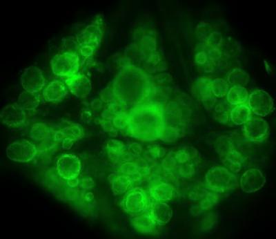
The topic of adult neurogenesis has been controversial over the past couple of decades due to a lack of strong evidence that new neurons are generated in humans after adolescence. However, the development of new technologies and experimental techniques – as well as an alarming rise in Alzheimer’s disease and other dementias1,2 – has brought adult neurogenesis back into the limelight.
There is a prevailing belief that neurogenesis – i.e. the growth and development of new neurons – halts once we reach adulthood. Indeed, most neurons are generated in the embryo, before birth.
Adult neurogenesis, however, refers to the continued generation of neurons from neural stem cells in the adult brain. These newborn cells eventually become indistinguishable from those born during embryonic development3.
Determining whether new neurons are continuously generated in the adult brain in humans, and how this process may be altered in both normal, ‘healthy’ aging and in neurodegenerative diseases such as Alzheimer’s, is an important question with potentially game-changing therapeutic potential.
In animal models, reductions in adult neurogenesis have been associated with a number of cognitive deficits including impairments in spatial memory, long-term memory and fear conditioning. Impaired neurogenesis has also been associated with a number of psychiatric disorders including depression, anxiety, addiction and schizophrenia. For example, patients with major depression have shown reduced levels of neurogenesis in the hippocampus, while antidepressants are known to increase neurogenesis in this region – an action which may be important in their therapeutic action4,5.
The hippocampus is involved in the consolidation of memories to other brain regions as well as in memory retrieval, and is one of the most affected areas in Alzheimer’s disease6. The dentate gyrus – a subsection of the hippocampus – plays a key role in memory retrieval, and is one of only two regions where the addition of new neurons has been observed throughout life in mammals. The other is the subventricular zone of the lateral ventricles, where the differentiation of new neurons plays a role in the sense of smell – although the latter is unlikely have any functional significance in humans7,8. Although adult neurogenesis in these two regions has been observed rodents and monkeys9, neither has been reliably observed in humans.
A seminal study by Eriksson et al. in 199810 provided the first direct evidence of adult neurogenesis in the hippocampus of humans. The researchers were not, however, able to quantify the number of new neurons in the samples, nor did it provide any insights into the dynamics of adult neurogenesis.
More recently, a study in 201311 built upon research in the field by devising a method to retrospectively determine the birth date of cells in the hippocampus. They achieved this by measuring the concentration of 14C in the DNA. The theory behind this approach was that, due to above-ground nuclear bomb testing during the Cold War, atmospheric 14C levels were elevated during the time period from 1955-1963; since then, levels of 14C in the atmosphere have steadily declined. Much of that 14C has reacted with oxygen to form CO2 which has in turn been taken up by plants through photosynthesis before passing through the food chain into animals and humans. As a result, atmospheric 14C levels are mirrored in human genomic DNA, as the 14C is incorporated into chromosomes during cell division at a concentration corresponding to that in the atmosphere at the time – thus providing a method of determining the cells’ date of birth. Effectively, a radioactive timestamp.
By measuring concentrations of 14C in human post-mortem hippocampal cells from subjects aged 19-92, the researchers could devise a model to calculate the rate of cell turnover which suggested that neurogenesis may indeed persist throughout adulthood. However, this was only a model – actual evidence of immature or newborn neurons being incorporated into adult brains in humans was still lacking.
Last year, a study led by Sorrells et al.12 attracted a lot of attention in the neuroscience community after publishing findings in which the authors failed to show any evidence of new neurons in post-mortem brain samples from adult humans. The research looked at the numbers of neural precursor cells and immature neurons in human port-mortem samples and concluded that neurogenesis in the hippocampus was most significant within the first year of life, before declining rapidly throughout childhood until eventually halting entirely in the brains of adults.
A paper published in Nature earlier this year, however, found quite the contrary.
The research13, led by Moreno-Jiménez et al. at the Department of Molecular Neuropathology, Universidad Autónoma de Madrid, Spain, aimed to investigate the extent of adult neurogenesis in the human hippocampus using post-mortem brain samples obtained from both healthy individuals and Alzheimer’s disease patients.
The research13, led by Moreno-Jiménez et al. at the Department of Molecular Neuropathology, Universidad Autónoma de Madrid, Spain, aimed to investigate the extent of adult neurogenesis in the human hippocampus using post-mortem brain samples obtained from both healthy individuals and Alzheimer’s disease patients.
Previous studies have shown both increases14 and decreases15 in neurogenesis in post-mortem tissue derived from patients with Alzheimer’s disease. Moreno-Jiménez and her team, however, sought to achieve more definitive results by improving upon the tissue-processing methods used in previous studies; proposing that previous failures to show direct evidence of adult neurogenesis in humans may actually be down to the experimental methods used in those studies.
Thus, by using state-of-the-art methods to tightly control the conditions by which the brain samples were obtained and prepared, the team achieved a greater sensitivity allowing them to identify thousands of immature neurons in the dentate gyrus of hippocampi from 13 neurologically healthy human subjects and 45 subjects with Alzheimer’s disease. While the numbers of new neurons declined throughout life in both cohorts – suggesting a natural age-related decline in neurogenesis – immature neurons were identified right through to the ninth decade of life.
The researchers identified newborn neurons using fluorescent antibodies, allowing them to visualise a specific marker of neuroblasts (neuronal precursor cells which will later develop into neurons) called doublecortin (DCX) (Figure 1). Moreno-Jiménez et al. reported a several-fold higher density of these types of immature cells in the dentate gyrus than has been reported in previous studies – most likely owing to the enhanced tissue preparation methods. The team also demonstrated the co-existence of several other neuroblast-associated markers, confirming that the cells identified were in fact immature neurons.
 |
| Figure 1: Confocal microscopy images showing (1) the entire hippocampus, (2) the dentate gyrus (DG) showing the abundant presence of DCX+ immature neurons (yellow triangles) and (3) a high-power-magnification image of one of these cells. Moreno-Jiménez et al. (2019) |
 |
Figure 2: The density of DCX+ neural precursor cells
is lower in Alzheimer’s disease patients compared with controls and declines with age, with counts dropping further as patients progress through the six Braak stages of Alzheimer’s disease. Moreno-Jiménez et al. (2019) |
Even more interesting is that the reductions in adult neurogenesis were also detected in the early stages of the disease – before the presence of neurofibrillary tangles or senile plaques thought to underlie the pathophysiology of Alzheimer’s.
It is possible therefore that detection of impairments in adult hippocampal neurogenesis using non-invasive methods may serve as an early biomarker for the disease, and that therapeutic targeting of cellular pathways underlying neurogenesis in the hippocampus could potentially mitigate the memory deficits characteristic of Alzheimer’s disease and other dementias.
A new study published in Cell Stem Cell this week16 by researchers at the University of Illinois in Chicago reported similar findings in post-mortem samples taken from a cohort of 18 patients with Alzheimer’s disease and other mild cognitive impairments, aged between 79 and 99 years old.
It is possible therefore that detection of impairments in adult hippocampal neurogenesis using non-invasive methods may serve as an early biomarker for the disease, and that therapeutic targeting of cellular pathways underlying neurogenesis in the hippocampus could potentially mitigate the memory deficits characteristic of Alzheimer’s disease and other dementias.
A new study published in Cell Stem Cell this week16 by researchers at the University of Illinois in Chicago reported similar findings in post-mortem samples taken from a cohort of 18 patients with Alzheimer’s disease and other mild cognitive impairments, aged between 79 and 99 years old.
Similarly, the researchers investigated the levels of DCX-positive neuroblasts in the samples, finding that these neural precursor cells, among other markers of developing neurons, were present in the hippocampus of brain tissue taken from patients well into their 90’s.
As well as supporting Moreno-Jiménez et al.’s conclusions that a decline in neurogenesis may be an important underlying feature of Alzheimer’s – potentially driving the decline in cognitive ability from the early stages of the disease – the study goes further by showing that higher counts of DCX-positive neuroblasts are associated with higher scores in measures of cognitive ability and an overall better clinical diagnosis.
It is worth noting, however, that while the results of these studies strongly support the general consensus among neuroscientists that neurogenesis does indeed persist throughout life in the hippocampus of humans, the existence of these neuroblast markers does not necessarily prove that these precursor cells all develop into fully mature neurons – studies have shown that newborn neurons that do not complete cell maturation and integration processes are eliminated during adult neurogenesis in mice16. Nonetheless, the results are in stark contrast to previous research which has generally found a decline in neuroblast numbers in the adult hippocampus throughout life11. These findings therefore provide crucial new evidence – reigniting the age-old debate about the relevance of adult neurogenesis in humans.
Since adult-born neurons have been found to contribute to learning and memory in animal models, it is quite feasible that this may also be true in humans. Adult neurogenesis may indeed be a fundamental part of learning and memory, with impairments in the addition, maturation or survival of newborn neurons potentially leading to memory deficits such as those seen in Alzheimer’s disease and other forms of dementia.
While these findings are the first to present direct evidence suggesting that neurogenesis does indeed continue throughout life in humans, research in the field is still in its infancy. Nevertheless, the results raise a host of questions regarding their potential to pave the way for new treatments for neurodegenerative diseases. What if we could isolate and therapeutically target a particular pathway or pathways involved in the regulation of adult neurogenesis in the hippocampus? Could this lead to improvements in memory in those suffering from Alzheimer’s and other dementias, potentially slowing or preventing the progression of the disease – or perhaps even offer a way of staving off the natural decline in memory due to ageing in healthy individuals? Future research will inevitably seek to answer these questions.
On a final note – numerous studies have shown that both exercise and sleep can significantly increase neurogenesis in mice, as well as stave off the natural decline due to aging17–20. Yet another reason to get more of both!
References:
1. Winblad, B. et al. Defeating Alzheimer’s disease and other dementias: a priority for European science and society. The Lancet Neurology 15, 455–532 (2016).
2. Chan, K. Y. et al. Epidemiology of Alzheimer’s disease and other forms of dementia in China, 1990–2010: a systematic review and analysis. The Lancet 381, 2016–2023 (2013).
3. Laplagne, D. A. et al. Functional Convergence of Neurons Generated in the Developing and Adult Hippocampus. PLOS Biology 4, e409 (2006).
4. Gonçalves, J. T., Schafer, S. T. & Gage, F. H. Adult Neurogenesis in the Hippocampus: From Stem Cells to Behavior. Cell 167, 897–914 (2016).
5. Miller, B. R. & Hen, R. The current state of the neurogenic theory of depression and anxiety. Current Opinion in Neurobiology 30, 51–58 (2015).
6. Braak, H. & Braak, E. [Morphology of Alzheimer disease]. Fortschr Med 108, 621–624 (1990).
7. Bergmann, O. et al. The Age of Olfactory Bulb Neurons in Humans. Neuron 74, 634–639 (2012).
8. Wang, C. et al. Identification and characterization of neuroblasts in the subventricular zone and rostral migratory stream of the adult human brain. Cell Research 21, 1534–1550 (2011).
9. Kornack, D. R. & Rakic, P. Continuation of neurogenesis in the hippocampus of the adult macaque monkey. PNAS 96, 5768–5773 (1999).
10. Eriksson, P. S. et al. Neurogenesis in the adult human hippocampus. Nature Medicine 4, 1313–1317 (1998).
11. Spalding, K. L. et al. Dynamics of Hippocampal Neurogenesis in Adult Humans. Cell 153, 1219–1227 (2013).
12. Sorrells, S. F. et al. Human hippocampal neurogenesis drops sharply in children to undetectable levels in adults. Nature 555, 377–381 (2018).
13. Moreno-Jiménez, E. P. et al. Adult hippocampal neurogenesis is abundant in neurologically healthy subjects and drops sharply in patients with Alzheimer’s disease. Nature Medicine 25, 554 (2019).
14. Jin, K. et al. Increased hippocampal neurogenesis in Alzheimer’s disease. PNAS 101, 343–347 (2004).
15. Li, B. et al. Failure of Neuronal Maturation in Alzheimer Disease Dentate Gyrus. J Neuropathol Exp Neurol 67, 78–84 (2008).
16. Tobin, M. K. et al. Human Hippocampal Neurogenesis Persists in Aged Adults and Alzheimer’s Disease Patients. Cell Stem Cell (2019). doi:10.1016/j.stem.2019.05.003
17. Sierra, A. et al. Microglia Shape Adult Hippocampal Neurogenesis through Apoptosis-Coupled Phagocytosis. Cell Stem Cell 7, 483–495 (2010).
18. Kreutzmann, J. C., Havekes, R., Abel, T. & Meerlo, P. Sleep deprivation and hippocampal vulnerability: changes in neuronal plasticity, neurogenesis and cognitive function. Neuroscience 309, 173–190 (2015).
19. Lucassen, P. J. et al. Regulation of adult neurogenesis by stress, sleep disruption, exercise and inflammation: Implications for depression and antidepressant action. European Neuropsychopharmacology 20, 1–17 (2010).
20. Vukovic, J., Colditz, M. J., Blackmore, D. G., Ruitenberg, M. J. & Bartlett, P. F. Microglia Modulate Hippocampal Neural Precursor Activity in Response to Exercise and Aging. J. Neurosci. 32, 6435–6443 (2012).
21. Clemenson, G. D. et al. Enrichment rescues contextual discrimination deficit associated with immediate shock. Hippocampus 25, 385–392 (2015).
Twitter image by Jason Snyder.
Twitter image by Jason Snyder.